Esclerosis Multiple Mri : Mri Brain With Contrast Multiple Sclerosis Mri Scan Images Mri Brain Multiple Sclerosis Multiple Sclerosis Tattoo - Multiple sclerosis (ms) is a common central nervous system (cns) disease characterised pathologically by the development of multifocal inflammatory demyelinating white matter lesions.
Esclerosis Multiple Mri : Mri Brain With Contrast Multiple Sclerosis Mri Scan Images Mri Brain Multiple Sclerosis Multiple Sclerosis Tattoo - Multiple sclerosis (ms) is a common central nervous system (cns) disease characterised pathologically by the development of multifocal inflammatory demyelinating white matter lesions.. Frederik barkhof and robin smithuis. Multiple sclerosis, or ms, affects roughly 2.5 million people around the world. Multiple sclerosis (ms) is a chronic inflammatory and neurodegenerative disease characterized. Esclerosis múltiple técnicas de diagnóstico neurológico imagen por resonancia magnética. Widespread use of mri (magnetic resonance imaging) has revolutionized the ability to diagnose multiple sclerosis.
And the exclusion of alternative diagnoses. Usually, mri is the only imaging modality needed for imaging patients with ms, and it far surpasses all other tests. Multiple sclerosis, or ms, affects roughly 2.5 million people around the world. Frederik barkhof and robin smithuis. Lo que el radiólogo debe conocer e informar.
Para Que Sirve La Resonancia Magnetica En Esclerosis Multiple from www.emyaccion.com Its high sensitivity for the evaluation of inflammatory and neurodegenerative processes in the brain and spinal cord has made it the most commonly used technique for the evaluation of patients with ms. Amsterdam university medical center and university college london and alrijne hospital leiderdorp, the netherlands. Lo que el radiólogo debe conocer e informar. Preferred examination radiologically, mri has revolutionized the investigation, diagnosis, and even the treatment of ms. The most determining test, capable of detecting plaques or scars that could be caused by multiple sclerosis, is magnetic resonance imaging (mri). Magnetic resonance imaging (mri) is one of the most important and most commonly used tools for diagnosing and monitoring multiple sclerosis (ms). Owing to its ability to depict the pathologic features of multiple sclerosis (ms) in exquisite detail, conventional magnetic resonance (mr) imaging has become an established tool in the diagnosis of this disease and in monitoring its evolution. There are multiple lesions in the spinal cord.
About 85% of individuals with ms are diagnosed.
Widespread use of mri (magnetic resonance imaging) has revolutionized the ability to diagnose multiple sclerosis. Optic nerve evaluation within normal limits. Magnetic resonance imaging (mri) plays a crucial role in multiple sclerosis (ms) diagnosis, disease monitoring, prognostication, and research. There are multiple lesions in the spinal cord. Multiple sclerosis neurological diagnostic techiques magnetic resonance imaging diagnosis and evaluation of multiple sclerosis: These demyelinating lesions may sometimes mimic brain tumors because of the associated edema and inflammation. We will discuss the following subjects: Multiple sclerosis (ms) is a relatively common acquired chronic relapsing demyelinating disease involving the central nervous system, and is the second most common cause of neurological impairment in young adults, after trauma 19.characteristically, and by definition, multiple sclerosis is disseminated not only in space (i.e. Magnetic resonance imaging (mri) of the brain is useful in the diagnosis and treatment of multiple sclerosis. An mri scan is abnormal in more than 95% of people recently diagnosed with ms. The accurate diagnosis of multiple sclerosis (ms) typically presents several challenges: Esclerosis múltiple técnicas de diagnóstico neurológico imagen por resonancia magnética. Lo que el radiólogo debe conocer e informar.
Magnetic resonance imaging (mri) is one of the most important and most commonly used tools for diagnosing and monitoring multiple sclerosis (ms). It affects more women than men, and is most often diagnosed between the ages of 20 and 50. About 85% of individuals with ms are diagnosed. The cns includes the brain, spinal cord, and optic nerves. But abnormal mri results do not always mean that you have ms.
Balo Concentric Sclerosis Radiology Reference Article Radiopaedia Org from prod-images-static.radiopaedia.org An mri scan is abnormal in more than 95% of people recently diagnosed with ms. Abnormalities show up on scans from many illnesses other than ms. Conventional magnetic resonance imaging (mri) has routinely been used to improve the accuracy of multiple sclerosis (ms) diagnosis and prognosis. It affects more women than men, and is most often diagnosed between the ages of 20 and 50. Usually, mri is the only imaging modality needed for imaging patients with ms, and it far surpasses all other tests. Its high sensitivity for the evaluation of inflammatory and neurodegenerative processes in the brain and spinal cord has made it the most commonly used technique for the evaluation of patients with ms. To describe the factors that are associated with gadolinium enhancement on mri in patients with multiple sclerosis (ms) and symptoms of relapse. There are multiple lesions in the spinal cord.
An mri scan is abnormal in more than 95% of people recently diagnosed with ms.
Amsterdam university medical center and university college london and alrijne hospital leiderdorp, the netherlands. Esclerosis múltiple técnicas de diagnóstico neurológico imagen por resonancia magnética. Multiple sclerosis (ms) is a chronic inflammatory and neurodegenerative disease characterized. Magnetic resonance imaging (mri) has developed into the most important tool for the diagnosis and monitoring of multiple sclerosis (ms). Magnetic resonance imaging (mri) plays a crucial role in multiple sclerosis (ms) diagnosis, disease monitoring, prognostication, and research. Frederik barkhof and robin smithuis. Usually, mri is the only imaging modality needed for imaging patients with ms, and it far surpasses all other tests. Multiple sclerosis (ms) is a common central nervous system (cns) disease characterised pathologically by the development of multifocal inflammatory demyelinating white matter lesions. However, conventional mri measures, such as the use of lesion volume and. Mri and ms multiple sclerosis (ms) is a condition in which the body's immune system attacks the protective covering (myelin) surrounding the nerves of the central nervous system (cns). Optic nerve evaluation within normal limits. Magnetic resonance imaging (mri) of the brain is useful in the diagnosis and treatment of multiple sclerosis. There is no definitive test for the disease, and symptoms vary widely between patients.
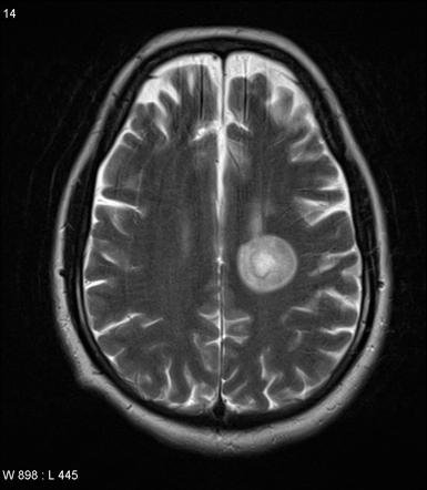
Multiple lesions in different regions of the brain) but also in time. Tumefactive multiple sclerosis is a term used to describe patients with established multiple sclerosis who develop large aggressive demyelinating lesions, similar/identical in appearance to those seen in sporadic tumefactive demyelinating lesions (tdl).tdl is now considered to be a separate entity, lying on a spectrum between multiple sclerosis and postinfectious demyelination/acute. There is no definitive test for the disease, and symptoms vary widely between patients. Mri and ms multiple sclerosis (ms) is a condition in which the body's immune system attacks the protective covering (myelin) surrounding the nerves of the central nervous system (cns). 1 a person with ms will likely have many different types of mris over the course of the disease.
Multiple Sclerosis The Role Of Mr Imaging American Journal Of Neuroradiology from www.ajnr.org These demyelinating lesions may sometimes mimic brain tumors because of the associated edema and inflammation. Amsterdam university medical center and university college london and alrijne hospital leiderdorp, the netherlands. But abnormal mri results do not always mean that you have ms. Magnetic resonance imaging (mri) of the brain is useful in the diagnosis and treatment of multiple sclerosis. Mri has made it possible to visualize and understand much more about the underlying pathology of the disease. It is the preferred imaging method to help establish a diagnosis of ms and to monitor the course of the disease. This article is an updated version of the 2013 article and focusses on the role of mri in the diagnosis of multiple sclerosis. We will discuss the following subjects:
Magnetic resonance imaging (mri) has developed into the most important tool for the diagnosis and monitoring of multiple sclerosis (ms).
These demyelinating lesions may sometimes mimic brain tumors because of the associated edema and inflammation. Multiple sclerosis (ms) is a common central nervous system (cns) disease characterised pathologically by the development of multifocal inflammatory demyelinating white matter lesions. 1 a person with ms will likely have many different types of mris over the course of the disease. The cns includes the brain, spinal cord, and optic nerves. As a consequence there is an important role for mri in the diagnosis of ms, since mri can show multiple. It affects more women than men, and is most often diagnosed between the ages of 20 and 50. Conventional magnetic resonance imaging (mri) has routinely been used to improve the accuracy of multiple sclerosis (ms) diagnosis and prognosis. Multiple sclerosis (ms) is the most common inflammatory. To describe the factors that are associated with gadolinium enhancement on mri in patients with multiple sclerosis (ms) and symptoms of relapse. Multiple lesions in different regions of the brain) but also in time. Optic nerve evaluation within normal limits. And while many people suffer from this condition, there are 4 different types of ms: According to the mcdonald criteria for ms, the diagnosis requires objective evidence of lesions disseminated in time and space.
1 a person with ms will likely have many different types of mris over the course of the disease esclerosis multiple. Topic overview an mri scan is the best way to locate multiple sclerosis (ms) lesions (also called plaques) in the brain or spinal cord.


Posting Komentar
0 Komentar